
In general, the goal of minimally invasive spine (MIS) surgery is to stabilize the vertebral bones and spinal joints and/or relieve pressure being applied to the spinal nerves — often a result of conditions such as spinal instability, bone spurs, herniated discs, scoliosis or spinal tumors.
As opposed to open spine surgery, minimally invasive surgical approaches can be faster, safer and require less recovery time. Because of the reduced trauma to the muscles and soft tissues (compared to open procedures), the potential benefits are:
- Better cosmetic results from smaller skin incisions (sometimes as small as several millimeters)
- Less blood loss from surgery
- Reduced risk of muscle damage, since less or no cutting of the muscle is required
- Reduced risk of infection and postoperative pain
- Faster recovery from surgery and less rehabilitation required
- Diminished reliance on pain medications after surgery
In addition, some MIS surgeries are performed as outpatient procedures and utilize only local anesthesia — so there is less risk for an adverse reaction to general anesthesia.
As with any surgical procedure, no matter how minimal, there are certain risks associated that include, but are not limited to:
- Possible adverse reaction to the anesthetic
- Unexpected blood loss during the procedure
- Localized infections, no matter how small the incision area
And, though uncommon, there is always a small chance that the initial MIS surgery cannot be completed, requiring either a second procedure or full open surgery.
Conditions Treated Using MIS Procedures
- Degenerative disc disease
- Herniated disc
- Lumbar spinal stenosis
- Spinal deformities such as scoliosis
- Spinal infections
- Spinal instability including spondylolisthesis
- Vertebral compression fractures
- Spinal tumors
How Minimally Invasive Spine Surgery Works
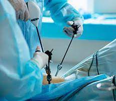
Because the spinal nerves, vertebrae and discs are located deep inside the body, any approach to gain access to the spinal area requires moving the muscle tissue out of the way. In general, this is facilitated by utilizing a small incision(s) and guiding instruments and/or microscopic video cameras through these incisions. Contrary to popular belief, lasers are very rarely used in MIS surgeries.
A number of methods can be used to minimize trauma during MIS surgery. Some of the more common techniques are outlined here.
Using a Tubular Retractor
This technique involves progressive dilation of the soft tissues, as opposed to cutting directly through the muscles. By using tubes to keep the muscles out of the way, the surgeon works through the incision without having to expose the area widely. Sometimes, the surgeon will also utilize an endoscope or microscope focused down the tube to assist with performing the surgery through a minimal access strategy. Once the procedure is complete, the tubular retractor can be removed, allowing the dilated tissues to come back together. Depending on the extent and type of surgery necessary, incisions can often be small.
Percutaneous Placement of Screws and Rods
Depending on the condition of the patient, it may be necessary to place instrumentation, such as rods and screws, to stabilize the spine or to immobilize the spine to facilitate fusion of the spinal bones. Traditional approaches for placement of screws requires extensive removal of muscle and other tissues from the surface of the spine.
However, percutaneous (meaning “through the skin”) placement typically involves inserting rods and screws through relatively small skin incisions without cutting or dissecting the underlying muscle. With the aid of x-ray images, guidewires are placed through the skin and into the spinal vertebrae along the desired paths for the screws. Then, screws are placed over the guidewires and follow the path of the wires. These screws have temporary extenders that extend outside of the skin and are subsequently removed after helping to guide passage of rods to connect and secure the screws. With the use of spinal navigation and robots, spinal instrumentation is being placed more safely and accurately.
Direct Lateral Access Routes
In some cases, especially those involving the lumbar spine, approaching the spine from the side of the body results in reduced pain, due to the limited amount of muscle tissue blocking the way. This approach is typically performed with the patient on his or her side. Then, a tubular retractor docks on the side of the spine to enable access to the spine’s discs and bones.
Thoracoscopic Access Route
Depending on the patient’s condition, it may be necessary to access the front portions of the thoracic spine, located in the chest and surrounded by the heart and lungs. Traditional access approaches often involve opening the chest through large incisions that may also require removal of one or more ribs. However, thoracoscopic access relies on multiple small incisions, through which working ports and cameras can be inserted to facilitate surgery.
Common MIS Surgery Treatment Options
A number of specific techniques have been deployed for MIS surgery. Though the field continues to develop, the list below highlights some of the most common options.
Discectomy: Spinal discs are essentially elastic rings with soft material inside that serve as cushions between the vertebral bones. If the elastic ring becomes weakened, the soft tissue inside can extrude — or herniate — outside of the elastic ring. The herniated disc material can compress the nerves passing by, thus causing pain. If surgical treatment is recommended to trim or remove the herniated disc, it may be possible to perform this procedure with MIS surgery using tubular dilators and a microscope or endoscope.
Spinal decompression:Spinal stenosis, which is a narrowing of the vertebral canal, is a common condition that can result in compression of the nerves. This can produce a variety of symptoms, including pain, numbness and muscle weakness. If surgery is recommended, it may be possible to remove the bone and soft tissues causing the nerve compression through an MIS approach using tubular dilators and a microscope or endoscope. The more common decompressive procedures include laminectomy and foraminotomy.
Transforaminal Lumbar Interbody Fusion (TLIF): This is a MIS technique that is performed for patients with refractory mechanical low back and radicular pain associated with spondylolisthesis, degenerative disc disease and recurrent disc herniation. The procedure is performed from the back (posterior) with the patient on his or her stomach. Utilizing two small incisions, screws and rods are placed between two or more vertebral levels. The intervertebral disc is removed and a cage filled with bone is placed in that void with the goal of stabilizing the levels affected.
How Can You Make a Difference in the Future of Spine Care
For more than 30 years, the NREF has funded research and training to improve treatments like minimally invasive spine surgery.Learn more about the NREF and make a donation today.
Candidates for MIS Surgery
A doctor will be able to tell which MIS surgeries, if any, might be an option for treating a spinal condition. In some situations, MIS surgery may not be as safe or effective as traditional open surgery. If so, the doctor will be able to inform you about the relative risks and benefits. In addition, there are some conditions that are not truly treatable with MIS surgery.
Glossary of Select Spine-Related Surgical Terms
- Bone spur: Bony growth or rough edges of bone (a.k.a. osteophyte).
- Decompression: A surgical procedure performed to relieve pressure and alleviate pain caused by the impingement of bone and/or disc material on the spinal cord or nerves.
- Disc degeneration: Degeneration or wearing out of a disc. A disc in the spine may deteriorate or wear out over time. A deteriorated disc may or may not cause pain.
- Discectomy: The surgical removal of part or all of an intervertebral disc, performed to relieve pressure on a nerve root or the spinal cord.
- Excision: Removal by cutting away material, as in removing a disc.
- Facet: A posterior structure of a vertebra which articulates (joins) with a facet of an adjacent vertebra to form a facet joint that allows motion in the spinal column. Each vertebra has a right and left superior (upper) facet and a right and left inferior (lower) facet.
- Foramen: A normal occurring opening or passage in the vertebrae of the spine through which the spinal nerve roots travel.
- Foraminotomy: Surgical opening or enlargement of the bony opening traversed by a nerve root as it leaves the spinal canal, to help increase space for that nerve.
- Herniated disc: A condition, also known as a slipped or ruptured disc, in which the gelatinous core material of a disc bulges out of position and puts painful pressure on surrounding nerve roots.
- Intervertebral foramen: An opening between vertebrae through which nerves leave the spine and extend to other parts of the body. Also known as neural foramen.
- Kyphosis: A condition in which the upper back curves forward, sometimes leading to the appearance of a hump in the back. Kyphosis may result from years of poor posture, spine fractures associated with osteoporosis, trauma or developmental problems.
- Lamina: The flattened or arched part of the vertebral arch, forming the roof of the spinal canal.
- Laminectomy: Surgical removal of the rear part of a vertebra in order to gain access to the spinal cord or nerve roots, to remove tumors, to treat injuries to the spine or to relieve pressure on a nerve root.
- Laminotomy: An opening made in a lamina, to relieve pressure on the nerve roots. As opposed to laminectomy (where the entire lamina is removed), a laminotomy typically involves removal of just half the lamina (the side where a patient is having symptoms).
- Lordosis: Lordotic curves refer to the inward curve of the lumbar spine. In some patients, this may represent a spinal deformity, also called swayback, which occurs when the lower back curves inward more than normal. Pathologic or excessive lordosis may be caused by osteoporosis or spondylolisthesis. Obesity, congenital disorders or overcompensation for kyphosis may contribute to this condition.
- Medial facetectomy: A procedure in which a part of the facet is removed to increase space in the spinal canal.
- Nerve roots: The initial portion of a spinal nerve; the nerve root is an extension of the central nervous system that begins at the spinal canal and ends in the extremities (fingers, toes). Its purpose is to send sensory information from the extremity to the brain and motor commands from the brain to the extremity.
- Pedicle: The bony part of each side of the neural arch of a vertebra that connects the lamina (back part) with the vertebral body (front part).
- Percutaneous: Effected, occurring or performed through the skin.
- Pseudarthrosis: The movement of a bone at the location of a fracture or a fusion resulting from inadequate healing of the fracture or failure of the fusion to mature properly. This can also result from a developmental failure.
- Scoliosis: Lateral (sideways) curvature of the spine.
- Spinal stenosis: Abnormal narrowing of the vertebral column that may result in pressure on the spinal cord, spinal sac or nerve roots arising from the spinal cord.
- Spinous process: A slender projection of bone from the back of a vertebra to which muscles and ligaments are attached.
- Spondylitis: Inflammation of vertebrae.
- Spondylolisthesis: The forward displacement of one vertebra on another.
- Spondylosis: Degenerative changes in the spine, most commonly affecting the intervertebral discs as well as the facet joints.
Device Technology
- Endoscope: A thin, fiberoptic tube with a light and lens, used to examine the interior of the patient’s body; provides minimally invasive access for diagnostic and surgical procedures.
- Fluoroscope: An imaging device that uses x-rays to view internal body structures on a screen, intraoperatively.
- Laparoscope: An instrument that enables visualization of specific structures within the body. A small surgical incision is made through which the laparoscope is placed. An array of tubes can be guided through the same incision, or other small incisions, permitting the use of probes and other instruments.
- Minimally invasive tubular retractor (MITR): Muscle-splitting technology first introduced in 1995 in conjunction with microendoscopic discectomy. The tubular retractor is used to create a tunnel down to the spinal column and can come in a variety of sizes, even as small as 1.4 cm in diameter (about 1/2 of an inch). A “muscle splitting” approach is employed, in which the tubular retractor is passed through a tunnel in the muscles of the back, rather than stripping the muscles away from the spine, as is done in open procedures. This approach limits damage to the muscles around the spine and decreases blood loss during surgery.
- Portals: Devices that provide a passage through which the surgeon operates during endoscopic procedures. After the incision is made, dilators are used to reach the area of the spine that the surgeon is working on. Fluoroscopy is used to locate the right level at time of surgery. During the procedure, instruments are used to continue the dissection through the portal. When the portal is removed, all the tissue falls back into place. In order to avoid damaging the tissue by moving instruments in and out of the passage, the portal or tubular retractor is placed into the incision to hold the tissue apart and left in place throughout the procedure. There are open and sealed portals. The portals used in the thoracic spine are usually 11 to 12 mm, while portals used in the abdominal cavity tend to be larger. All of the instruments and implants must be designed to fit through these small passages and perform surgical functions, once they reach the site.
Spinal Fusion
Spinal fusion is an operation that creates a solid union between two or more vertebrae. This procedure may assist in strengthening and stabilizing the spine and may thereby help to alleviate severe and chronic back pain.
Almost all of the surgical treatment options for fusing the spine involve placement of a bone graft between the vertebrae. Bone grafts may be taken from the hip or from another bone in the same patient (autograft) or from a bone bank (allograft). Bone graft extenders and bone morphogenetic proteins (hormones that cause bone to grow inside the body) can also be used to reduce or eliminate the need for bone grafts.
Fusion may or may not involve use of supplemental hardware (instrumentation), such as plates, screws and cages. This fusing of the bone graft with the bones of the spine will provide a permanent union between those bones. Once that occurs, the hardware is no longer needed, but most patients prefer to leave the hardware in place rather than go through another surgery to remove it. Fusion can sometimes be performed via smaller incisions through MIS techniques. The use of advanced fluoroscopy, endoscopy and navigation has improved the accuracy of incisions and hardware placement, minimizing tissue trauma while enabling an MIS approach.
MIS Fusion Procedures
- Minimally Invasive Lateral Interbody Fusion
- Minimally Invasive Posterior Lumbar Interbody Fusion (PLIF)
- Minimally Invasive Transforaminal Lumbar Interbody Fusion (TLIF)
- Minimally Invasive Posterior Thoracic Fusion